Clinician-centered AI for ear care
See clearly.Decide
confidently.
We’re developing an AI copilot to help clinicians review ear images, understand visible findings, and assess possible pathology—with clinical judgment at the center.
Technology under development Patent pending
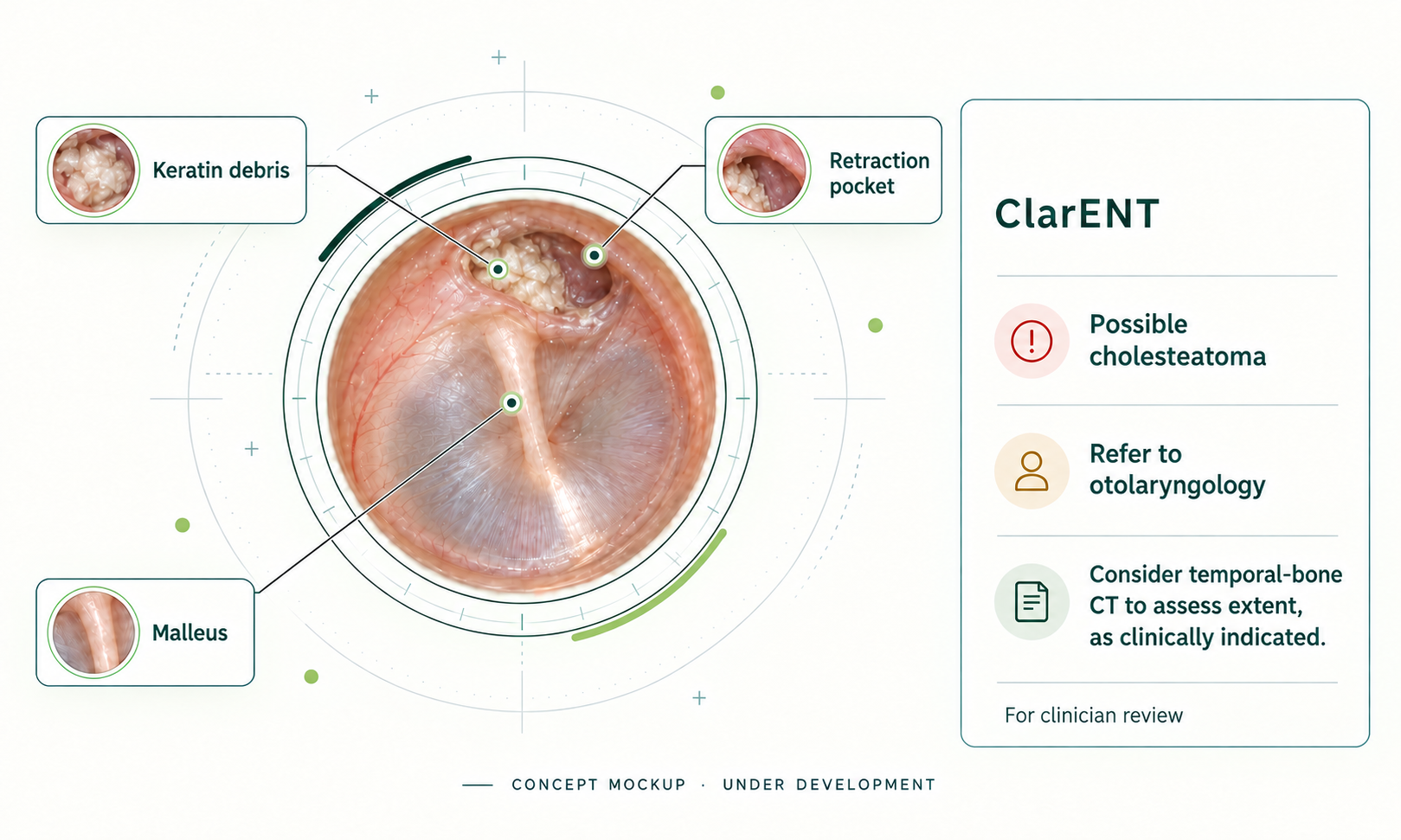
The otoscopy copilot
A clearer view of
the next step.
Our intended workflow connects image quality, visible findings, assessments of possible pathology, and clinician review.
Capture — start with a useful view
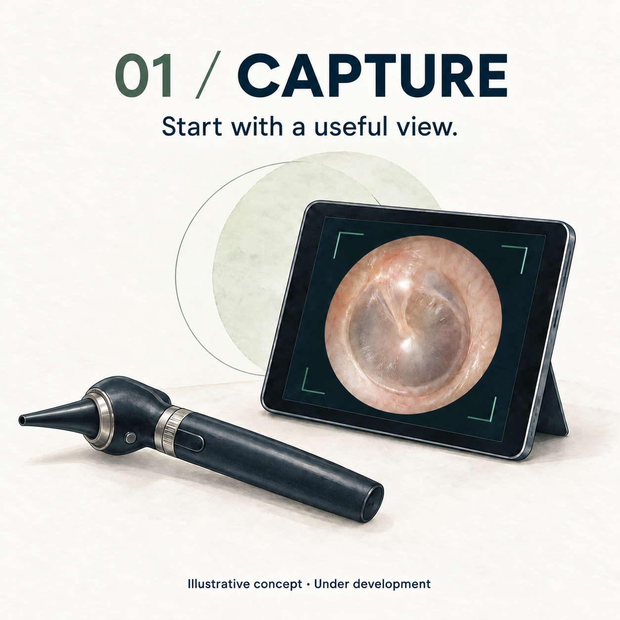
We’re developing image enhancement to reduce blur and improve poor illumination, with capture guidance to help clinicians obtain a clearer view of the tympanic membrane.
Understand — review visible findings
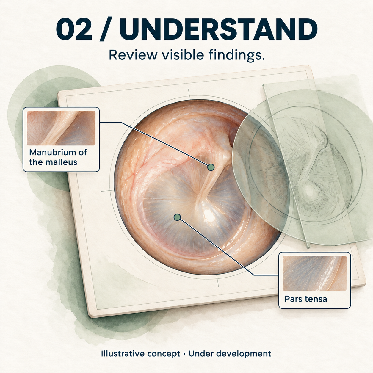
Review visible anatomy and localized findings, with assessments of possible pathology to support interpretation.
Consider — keep clinical judgment central

Bring the findings into clinical context as the clinician considers diagnosis, management, and referral.
Illustrative workflow concepts · Technology under development. Select an image to view it in detail.
A closer look · TM perforation
From visible findings to clinical review.
A possible eardrum perforation offers another example of how ClarENT could bring visible features and next-step considerations into one view for the clinician.
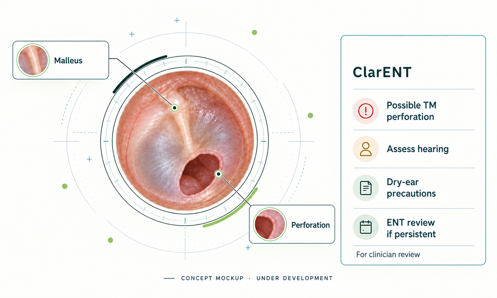
What’s visible
A possible tympanic membrane perforation is detected and flagged by the software. The clinician confirms the finding and reviews the case history and symptoms.
What the clinician considers
Assess hearing, discuss keeping water out of the ear, and arrange follow-up. Persistent perforation, hearing loss, or recurrent infections may prompt otolaryngology evaluation for repair.
Clinical context: AAO-HNS tympanoplasty clinical indicators. These clinical suggestions inform the example; care depends on the individual history and examination.
Planned feature
Follow healing over time.
We’re planning tools to measure tympanic membrane perforation size and track changes across follow-up visits. By showing reductions in size over time, ClarENT would help clinicians review healing progress alongside the patient’s symptoms and examination.
Built around clinical review
Clinical insight that
supports your judgment.
A useful image is the starting point. ClarENT is being developed to support interpretation while making uncertainty clear. Clinicians bring the patient’s history, examination, and their own judgment to every diagnosis and care decision.
Get to know ClarENTImprove visibility and guide a better capture before reviewing findings.
Keep possible explanations and uncertainty in view.
Support professional judgment without requiring a diagnostic label.
Specialist computer vision, controlled foundation-model reasoning, and clinical rules—brought together in a hybrid architecture under development.
The broader vision
More of the picture.
One connected approach.
Our future vision connects otoscopy, tympanometry, and audiometry in a coordinated ear-assessment workflow.
Otoscopy
Ear images, visible anatomy, and localized findings.
Tympanometry
Middle-ear measurements in a broader clinical context.
Audiometry
Hearing assessment as part of a more complete picture.
Multimodal integration is a future development direction. Graphics are illustrative, not patient results.
A few useful answers
Where we are.
Where we’re going.
What is ClarENT’s current stage?
ClarENT is under development. We’re seeking collaborators in clinical validation, machine learning, computer vision, and medical-device integration.
What image-quality features are in development?
We’re developing image enhancement to reduce visual blur and improve visibility in poorly illuminated otoscope images, alongside capture guidance to help clinicians obtain a clearer view of the tympanic membrane. The aim is to improve on the typical still image from a handheld digital otoscope and help clinicians recognize when a better capture is needed. These features are under development.
What is the Otoscopy Copilot intended to do?
The intended workflow supports review of image quality, tympanic-membrane visibility, visible anatomy, and localized findings, followed by assessments of possible pathology and support for clinical interpretation. Clinicians consider these assessments in the context of the patient’s history, examination, and other available findings.
Does ClarENT make clinical decisions or force a diagnosis?
ClarENT is being designed to provide assessments of possible pathology for clinician review. Diagnosis and care decisions remain with the clinician. The intended workflow allows more than one possible explanation or an inconclusive assessment; it does not require a diagnostic label when the available findings do not support one.
Is ClarENT intended to replace existing OEM software?
No, ClarENT is designed to integrate with existing OEM software to provide additional value and features including image enhancement and an AI copilot for clinician guidance.
Who can collaborate with ClarENT?
We welcome conversations with OEMs, medical-device teams, machine-learning and computer-vision specialists, clinicians, and researchers interested in development, validation, and integration. Contact us at info@clarentmedicalai.com.
Help shape what comes next
Better tools for ear care.
A shared ambition.
Tell us what you’re working on and where you see an opportunity to collaborate.
Explore collaboration- 01Clinical research and evaluation
- 02AI and computer vision development
- 03Medical-device and workflow integration